
 News
News
Acentech wins AN Best of Practice Award
Acentech is honored to be awarded an Editors’ Pick in The Architect’s Newspaper 2025 Best of Practice Awards in the Acoustic Consultant/Engineer category! This recognition highlights…
July 10, 2025
Here at Acentech’s Cambridge office, we are fortunate to be located down the street from world-class institutions doing research on hearing, a topic of interest in our work in acoustics. A pair of recent studies from researchers at Harvard Medical School, Boston Children’s Hospital, and Massachusetts Eye and Ear Infirmary have improved our understanding of the auditory system by examining structures embedded in or connected to each end of inner hair cells.
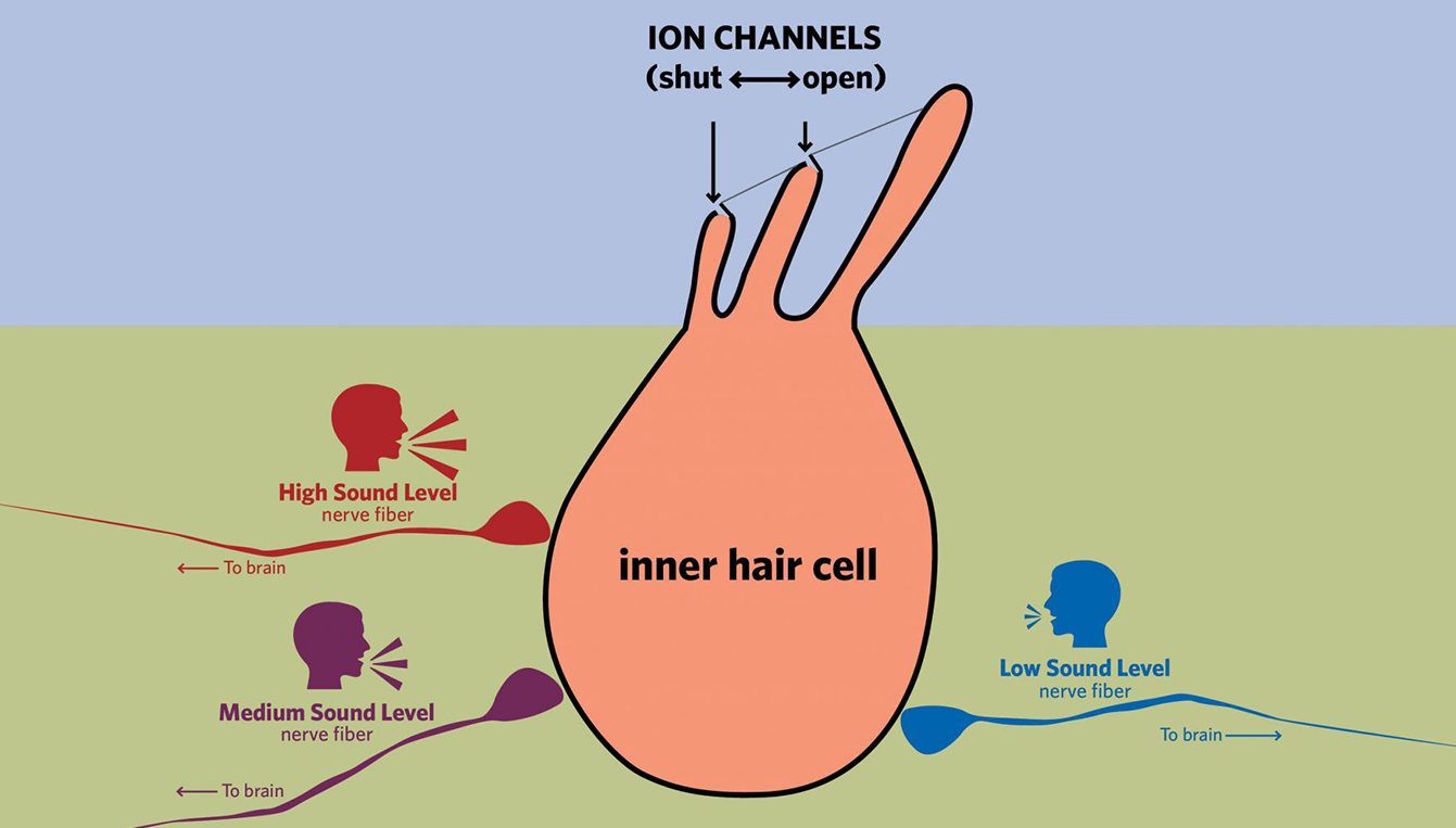
So, how does the auditory system actually work? In short, sound pressure in the air is collected by the outer ear and transformed into vibrations in the middle ear, which houses the smallest bones in the human body. These vibrations conducted through these bones are transmitted to fluid in the cochlea, a snail-shaped organ that separates the vibration by frequency, so it acts like a frequency analyzer. Motion of the fluid in the cochlea causes a trampoline-like region called the basilar membrane to vibrate, generating movement in inner hair cells, which function like microphones, changing mechanical energy into electrical signals. These electrical signals are then transmitted through connected auditory nerve fibers to the brain. These hair cells and nerve fibers are the focus of some of the most exciting hearing research today.
Bifeng Pan and colleagues from Boston Children’s Hospital, Harvard Medical School, Ohio State, and NIH looked at the ion channels at the tips of the hair cells that let ions into the cells. Ions are charged particles (also known as the electrolytes in your favorite sports drink) that carry electrical currents in biological systems. The ion channels at the tips of inner hair cells open and close in response to sound, switching between letting ions in to the cell and blocking them, like opening and closing the stopper in your bathroom sink. The movement of ions into the inner hair cell generates the electrical signal that is transmitted to the brain. For a long time, researchers have tried to grapple with a key question: what made up the ion channels and how were they shaped? The answer lied in a particular protein. Proteins are chains of amino acids and they twist and fold into interesting shapes depending on the order of the amino acids. This study focused on a particular protein TMC1 that was known to be involved in hearing. The researchers performed molecular experiments and computer simulations to determine the shape of the molecule. They also modified the protein to see its effects on inner hair cell function. The resulting experiments showed that the TMC1 protein made up the ion channels responsible for inner hair cell transduction. This discovery is important because it gives us a better sense of how the inner hair cell functions and it provides more information for potential therapies.
Another paper, authored by Brikha Shrestha and colleagues at Harvard Medical School and Massachusetts Eye and Ear Infirmary, examined the variation among nerve fibers that carry auditory signals from inner hair cells to the brain. Prior work has found that these fibers fall into three categories: one responsive to low sound levels, another responsive to moderate sound levels, and a third responsive to high sound levels. The high sound level fibers are important for understanding speech in noise and are unfortunately the most vulnerable to damage from noise exposure and aging. The current work found that different genes were expressed in each category and that this heterogeneous expression doesn’t occur until nerve fibers form synapses with the inner hair cells and receive stimulation from the inner hair cells. Knowing the molecular differences between these fibers provides clues about why these fibers function as they do and provides opportunities for genetic manipulation in mouse studies to learn more about the importance of each category. This research could potentially produce therapies for some types of congenital deafness.
M. Charles Liberman, Ph.D. of Harvard Medical School and Mass Eye and Ear, was kind enough to offer this additional insight into the findings of the research conducted by Shrestha et al.:
“The division of the auditory nerve fibers into three subtypes with different threshold sensitivities is key to the incredibly wide range of sound intensities over which our ears can operate. The discovery of the molecular basis for this functional subdivision provides us with powerful new tools to study fundamental aspects of hearing and hearing impairment.”
Although inner hair cells may be small, they carry big answers to the mysteries of the human ear. Thanks to diligent research, we are one step closer to helping individuals with debilitating hearing deficiencies.
News
Acentech is honored to be awarded an Editors’ Pick in The Architect’s Newspaper 2025 Best of Practice Awards in the Acoustic Consultant/Engineer category! This recognition highlights…
 News
News
Acentech worked on an astounding 30 projects that are being toured at the AIA25 Conference on Architecture in Boston next month! We had a great time…
 News
News
During the AIA25 Conference (June 4-7), Acentech will have a pop-up 3DListening studio at Boston Society for Architecture’s BSA Space at 290 Congress Street in Boston!…